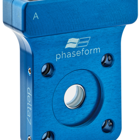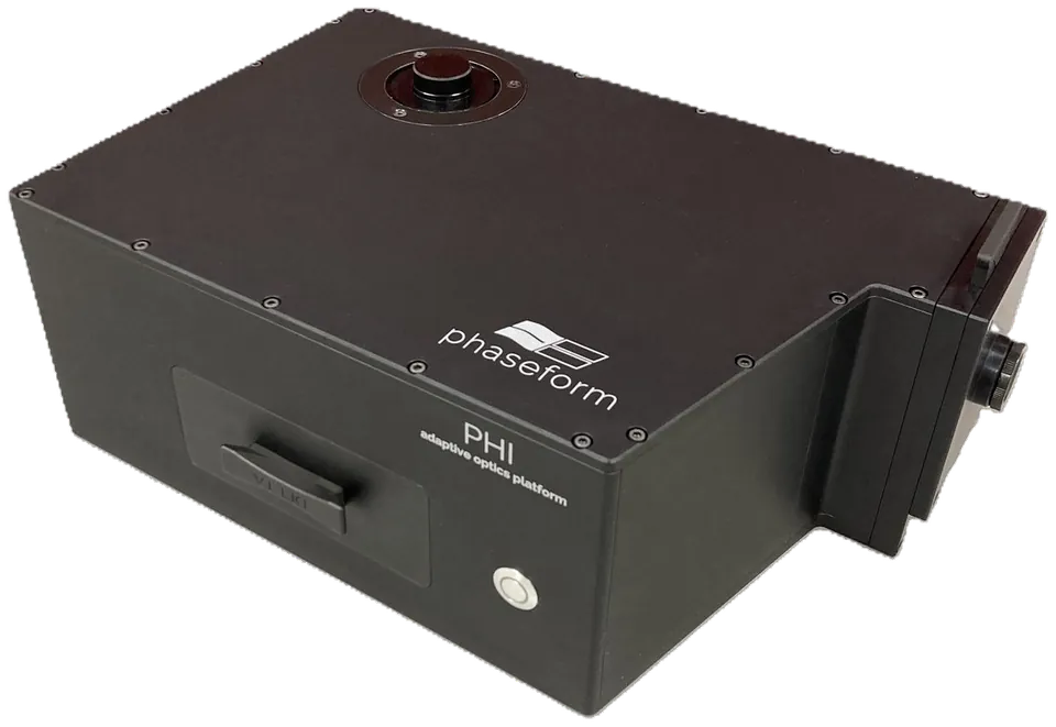

Description
 Adaptive optics through the camera port
- Plug-and-playAttaches to any standard C-mount camera port. Connect power and USB, and you are ready to go.
- ModularEasily insert or remove the DPP to switch between standard imaging and adaptive optics.
- Adaptive opticsCorrect up to the 7th radial order of Zernike modes. Automatically optimize image quality for thick, scattering tissue samples.
- Motionless 3D focusingIntegrated tunable lens at the conjugate pupil plane with up to 16 diopters of focusing range: a z-stack costs no stage motion, no settling time and no mechanical disturbance of the sample.
- Future-proofWorks with current and upcoming DPPs: swap in the one that suits your application, without replacing the platform.
- Stays on the portBetween experiments PHI runs in bypass at > 90 % average transmission, 450 to 1050 nm — it earns its place on the camera port whether or not you are correcting that day.
See what PHI does
Results from partner labs: aberration the sample introduces, deliberate wavefront shaping, and single-molecule localization.
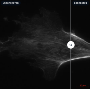
Correcting what the sample introduces
a wrong immersion medium — on a specimen, and on point sources — Life Imaging Center, University of Freiburg
Driving the wavefront on purpose — PSF shaping
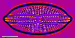
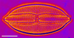
Single focal plane. 3D projection
Navicula lyra, 100× oil objective. Left is one focal plane; right is a projection through a z-stack acquired with PHI’s tunable lens: ~14 nm axial steps over ~3.4 µm of depth. The tunable lens sits at the conjugate pupil plane, so refocusing is electrical: the objective never moves. Scale bar 10 µm.ZEISS Axiovert 200M, Life Imaging Center, University of Freiburg. Courtesy of Dr. Roland Nitschke.
Single-molecule localization
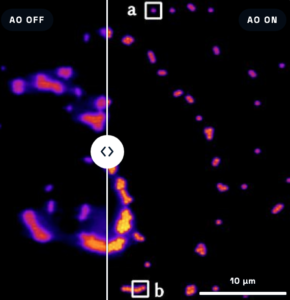
the run and the PSF underneath it — TU Wien, Biophysics Research Unit
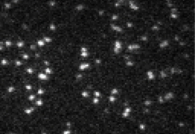
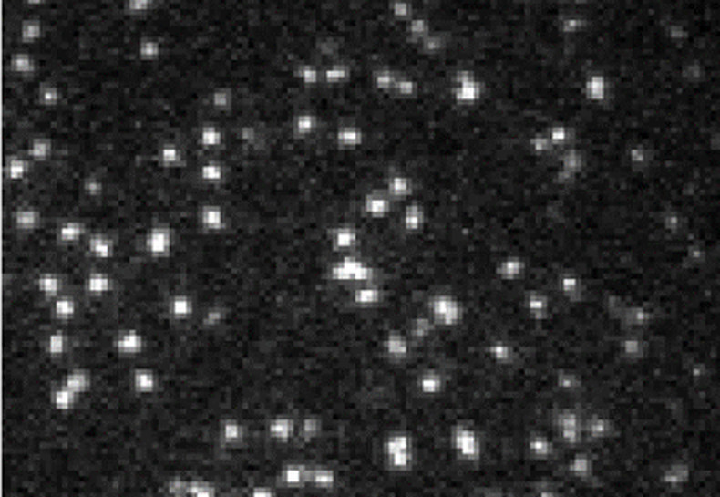
Without AO With AO
Mobile ATTO488-DOPE in a supported lipid bilayer under TIRF illumination, ZEISS Plan-Apochromat 100×/1.46. PHI corrects the sample-induced aberration while the localization run is acquiring.TU Wien, Institute of Applied Physics, Biophysics Research Unit. Courtesy of Prof. Gerhard Schütz and Dr. Mario Brameshuber.
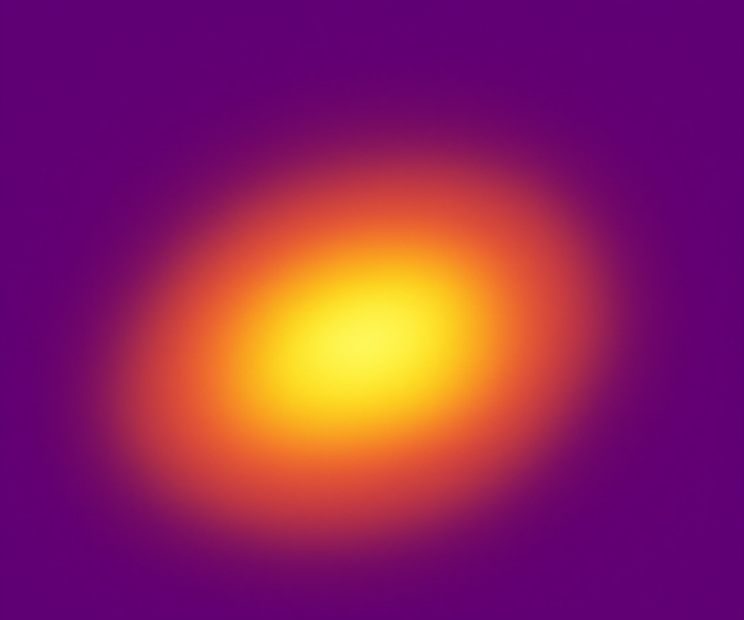
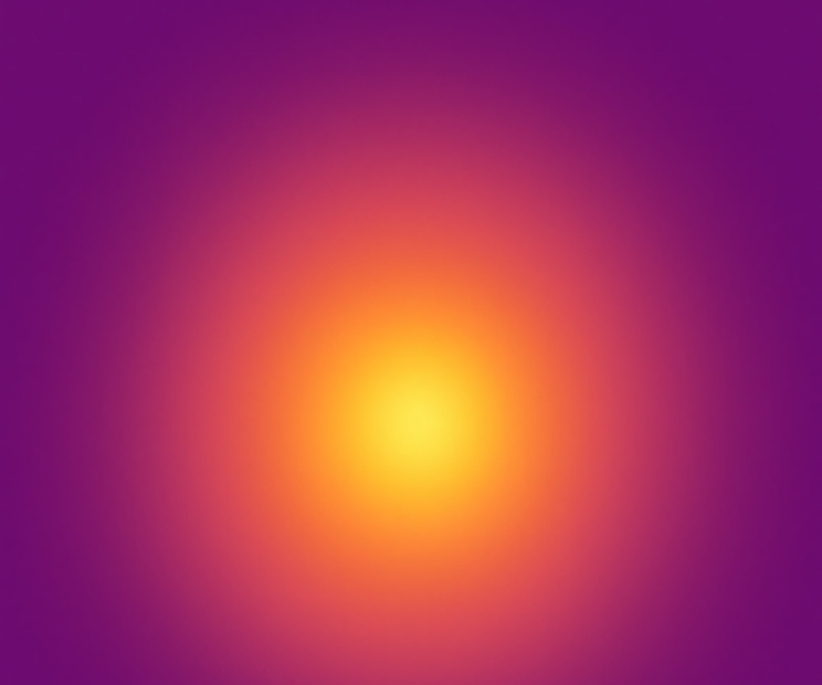
Aberrated. Corrected
The measured 2D point-spread function from the same single-molecule setup, both panels on the same scale. Localization accuracy depends on PSF symmetry: the aberrated PSF is elongated and tilted, the corrected one is round.TU Wien, Institute of Applied Physics, Biophysics Research Unit. Courtesy of Prof. Gerhard Schütz and Dr. Mario Brameshuber.
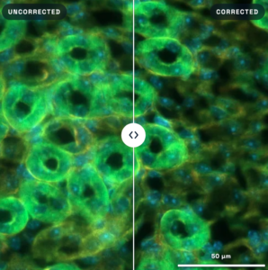
Thick tissue
the hardest case — aberration the sample itself introduces, varying across the field
Specifications
PHI at a glance
What it attaches to and what the camera port has to carry, what it does to the wavefront, what it needs to run — and, at the end, whether your own objective’s pupil lands inside the window.
Compatibility
| Microscope | Mounts onto any standard C-mount port of upright or inverted microscope frames Chain multiple C-mount devices: confocal & spinning-disk modules, structured illumination, etc. |
|---|---|
| Objective lens | Covers objectives with pupil diameters from 5 mm to 20 mm 5–11 mm using the Delta 7-10 · 11–20 mm using the Delta 7-20 |
| Camera | C-mount cameras supported F-mount version in preparation |
| Software | µManager, ScanImage, ThorImage, ZEN blue or the Python SDK Nikon NIS-Elements integration in preparation |
| Overall envelope (W × D × H) | 367.5 × 230.4 × 132 mm |
| Camera port (in) | C-mount female (internal thread) |
| Camera port (out) | C-mount male (external thread) |
| Weight | 8.3 kg |
Performance
Adaptive optics
- Maximum peak-to-valley of the corrected wavefronts: > 10 µm
- Maximum spatial frequency of the correction: 7th radial order of Zernike modes
- Response time < 40 ms
Remote focusing
- Tuning range up to 16 dpt
- Tuning precision 0.001 dpt
- Response time 3 ms
Optical transmission
- AO & remote-focusing mode ~ 75 % average, 450–1050 nm
- Bypass mode > 90 % average, 450–1050 nm
General
- Power & connectivity 12 V wall power, USB to PC
- Main applications spherical-aberration correction, deep-tissue imaging, remote focusing, 3D imaging
Mechanical envelope
The DPP exchange slot on the side panel and the insertable pinhole on the output side stay reachable while the unit is mounted: swap the Delta 7 without taking PHI off the microscope.
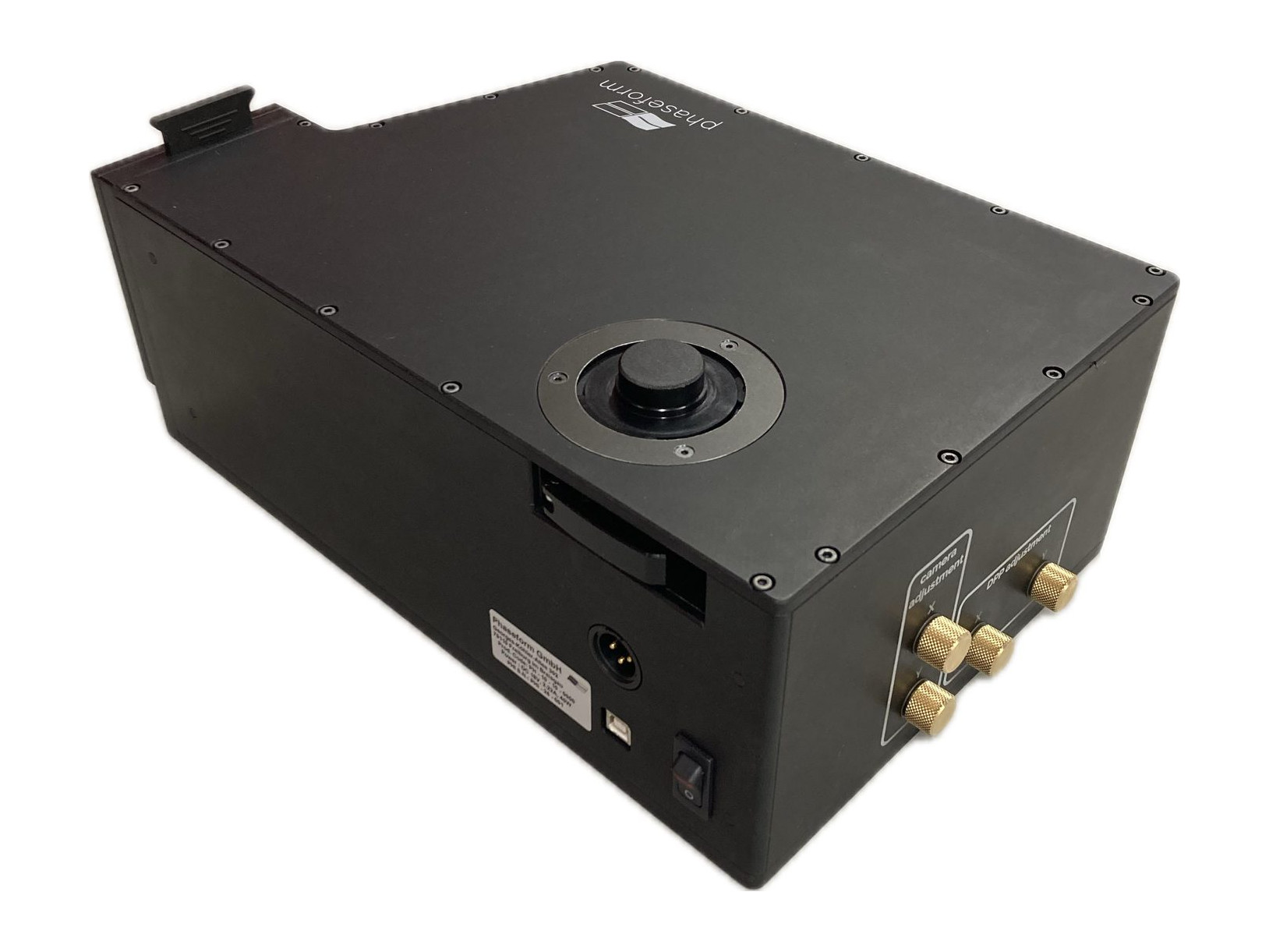
Camera release lever · DPP & camera adjustment nubs.
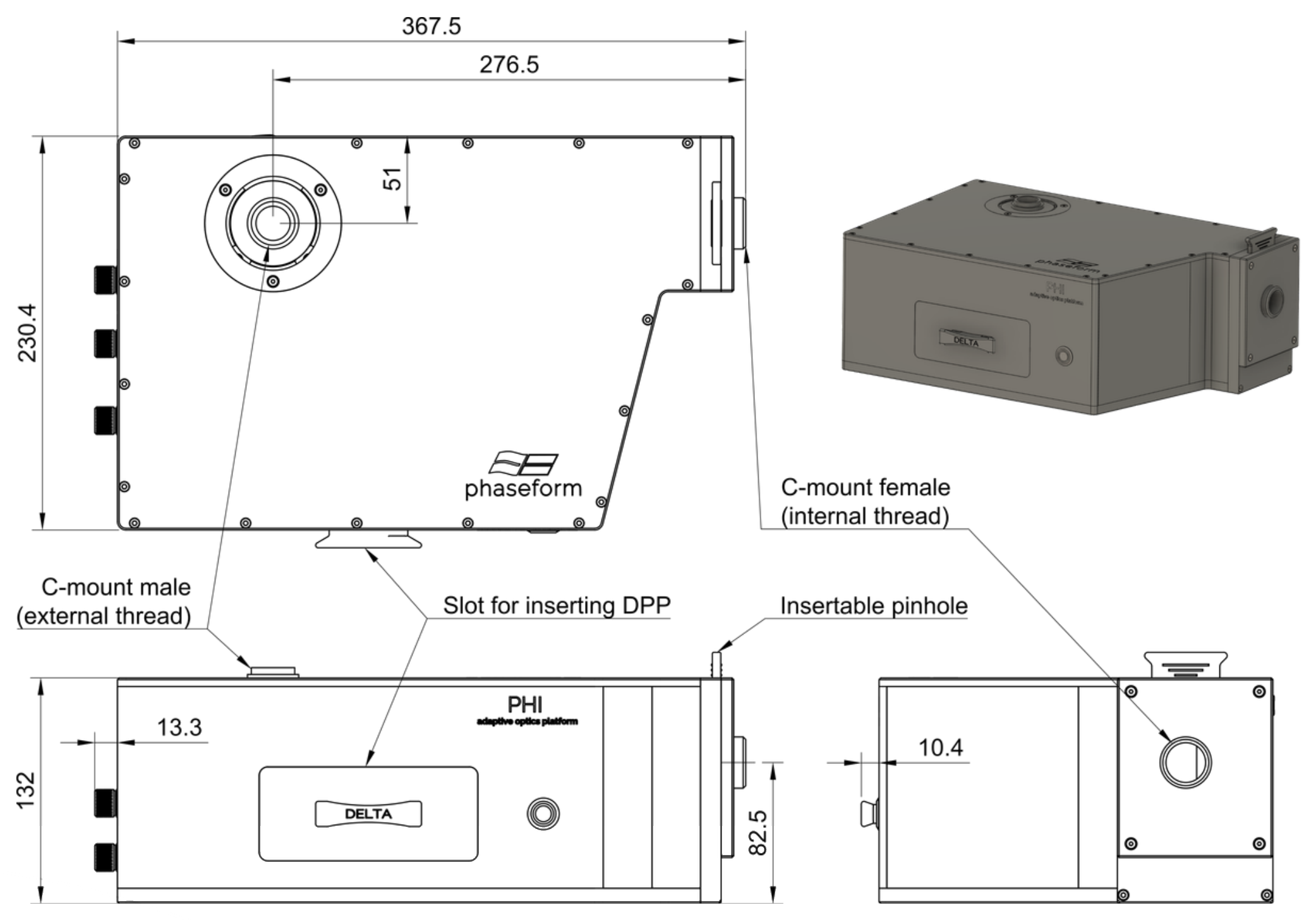
All dimensions in millimeters.
Objective lens
| Mag | 4× | 10× | 20× | 25× | 40× | 63× | 100× |
|---|---|---|---|---|---|---|---|
| Min NA | 0.2compatible | 0.25compatible | 0.4compatible | 0.7compatible | 0.8compatible | 1.2compatible | 1.25significantly reduced performance |
| Max NA | 0.28significantly reduced performance | 0.45compatible | 0.75compatible | 1.1compatible | 1.3compatible | 1.45compatible | 1.45compatible |
CompatibleSignificantly reduced performance
Typical NA range each magnification is sold in, against PHI’s 5–20 mm pupil window at a 200 mm tube lens; Olympus (180 mm) and ZEISS (165 mm) frames shift the result.